November 26, 2015 report
New way to use ultrasound allows for imaging live blood vessels with more clarity

(Medical Xpress)—A team of researchers affiliated with several institutions in France has developed a new way to create live images of blood vessels. In their paper published in the journal Nature, the team describes their new technique, the results they have managed to achieve and the ways the new technology might be used. Ben Cox and Paul Beard with University College London offer a News & Views piece on the work done by the team along with a historical perspective, in the same journal issue.
Doctors would really like to be able to watch blood flowing in any part of the body in real time—that would allow them to see blockages, tumors and a host of other medical problems in their patients. Unfortunately, that is still not possible, though ultrasound and light microscopy are constantly improving, leading perhaps to that ultimate goal. In this new effort, the researchers report that they have taken a step closer using an approach that involves combining ultrasound and microscopic bubbles injected into the bloodstream of a living rat.
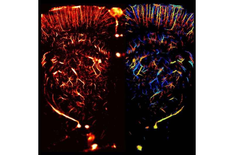
In their approach, extremely small gas bubbles, on the order of 2 micrometers, are injected into the bloodstream in a given area, such as an organ. Once they are inside the blood vessels, high frequency ultrasound waves are fired through the skin and into the area under study. When the waves hit the bubbles, some of the energy is bounced back. That energy is captured by an imaging system that operates at several thousand frames per second, allowing for distinguishing between echoes and bubble data. As each individual bubble moves, its progress is captured—putting all the individual images together allows for creating a single image of the entire vessel or network of vessels.
The researchers report that it took just ten seconds to build such an image of the blood vessels in a rat's brain, with a resolution of 10 micrometers—the best that has ever been achieved using any technique.
The researchers believe their new technology will make it into the medical community, helping doctors not only find problems and treat them, but to learn more about how they unfold. One such example would be tracking the growth of new vessels surrounding a tumor, which could help predict how fast it is growing.
More information: Claudia Errico et al. Ultrafast ultrasound localization microscopy for deep super-resolution vascular imaging, Nature (2015). DOI: 10.1038/nature16066
Abstract
Non-invasive imaging deep into organs at microscopic scales remains an open quest in biomedical imaging. Although optical microscopy is still limited to surface imaging owing to optical wave diffusion and fast decorrelation in tissue, revolutionary approaches such as fluorescence photo-activated localization microscopy led to a striking increase in resolution by more than an order of magnitude in the last decade1. In contrast with optics, ultrasonic waves propagate deep into organs without losing their coherence and are much less affected by in vivo decorrelation processes. However, their resolution is impeded by the fundamental limits of diffraction, which impose a long-standing trade-off between resolution and penetration. This limits clinical and preclinical ultrasound imaging to a sub-millimetre scale. Here we demonstrate in vivo that ultrasound imaging at ultrafast frame rates (more than 500 frames per second) provides an analogue to optical localization microscopy by capturing the transient signal decorrelation of contrast agents—inert gas microbubbles. Ultrafast ultrasound localization microscopy allowed both non-invasive sub-wavelength structural imaging and haemodynamic quantification of rodent cerebral microvessels (less than ten micrometres in diameter) more than ten millimetres below the tissue surface, leading to transcranial whole-brain imaging within short acquisition times (tens of seconds). After intravenous injection, single echoes from individual microbubbles were detected through ultrafast imaging. Their localization, not limited by diffraction, was accumulated over 75,000 images, yielding 1,000,000 events per coronal plane and statistically independent pixels of ten micrometres in size. Precise temporal tracking of microbubble positions allowed us to extract accurately in-plane velocities of the blood flow with a large dynamic range (from one millimetre per second to several centimetres per second). These results pave the way for deep non-invasive microscopy in animals and humans using ultrasound. We anticipate that ultrafast ultrasound localization microscopy may become an invaluable tool for the fundamental understanding and diagnostics of various disease processes that modify the microvascular blood flow, such as cancer, stroke and arteriosclerosis.
© 2015 Medical Xpress


