New imaging technology enlightened by biomedical engineering

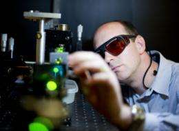
A Faculty of Engineering professor is conducting biomedical research that could have profound effects on medical imaging and the delivery of drugs by using light and ultrasound. Roger Zemp, a professor in the Department of Electrical and Computer Engineering, researches photo-acoustic imaging -- literally making vivid, high-resolution images of the "sound" of light.
The research is simple in principle. In photo-acoustic imaging a beam of light is aimed at a target beneath the skin—a tumour for example. When the light hits its target, it heats up slightly and the tissue expands, emitting acoustic waves. Technologists can use ultrasound equipment to make an image of the tissue as a result of its expansion.
“The light is absorbed and turns to heat, the tissue expands and we listen to that expansion,” Zemp explains, adding that the concept of photo-acoustic imaging was explored by Alexander Graham Bell more than 100 years ago, but has only recently been applied to biomedicine.
The benefits of photo-acoustic imaging are clear: it doesn’t carry with it the same radiation exposure that X-rays do, image quality is high and functional and molecular information can be extracted because of the high quality optical contrast. In the case of diagnostic tests like mammograms, photo-acoustic imaging could help in the early detection of cancer by providing doctors with more detailed, crisp images.
In cancer treatment photo-acoustic imaging helps because, “we can understand what is going on in a tumour a lot better,” said Zemp. “We can actually see, vessel by vessel, how much oxygen is in the blood.”
This is important because tumours often have hypotoxic regions—areas that are poorly vascularized and necrotic. Cells that experience hypoxia begin to behave in ways that make them resistant to some therapies, says Zemp, so having an accurate “map” of a tumour is valuable in treating it. Different variations of photoacoustic imaging systems are being developed: systems similar to diagnostic ultrasound systems found in hospitals, and specialized systems aiming to image capillary networks in real time.
Zemp’s team is also using photoacoustic imaging to image gene expression. Often, biologists want to observe whether a particular gene is being expressed in cells or in living subjects. They use ‘reporter genes’ which produce an optical signal to reveal the gene expression patterns. The problem is that such optical methods have poor penetration and spatial resolution in living subjects. Zemp and his colleagues are developing photoacoustic imaging techniques to visualize such gene expression patterns with high resolution at multiple-centimeter depths.
Zemp is working on other related research projects as well. In one, he has teamed up with postdoctoral fellow Robert Paproski and renowned oncologist Carol Cass. Cass is one of the pioneers in the field of nucleoside transport—the phenomena through which cells absorb drugs.
Because patients with pancreatic cancer are often unable to absorb drugs, Cass is looking for alternate delivery methods, or ways to cure the patients’ nucleoside transport deficiency.
Zemp and Paproski are investigating the use of micro bubbles to deliver genetic treatments or drugs. The microbubbles measure up to five microns in diameter, making them the size of a cell.
The idea is to first load the microbubbles with DNA that would encode cells with the ability to absorb drugs, deliver the bubbles to the pancreas, then use low-frequency ultrasound waves to pop them once the bubbles reach their target area.
Alternately, the same technique and technology could be used to deliver drugs directly to a tumour, minimizing side effects and enhancing the effect of the drug.
Zemp is also working on research to improve existing ultrasound and photo-acoustic imaging technology. In collaboration with a pediatric cardiologist, Zemp is looking for better ways to detect pulmonary hypertension. A telltale sign in diagnosing pulmonary hypertension is that the second sound in a heartbeat is split into two sounds.
“We envision having something like a stethoscope hooked up to a mobile phone and having an app that looks for the splitting of that second heart sound,” he said.
He is also working with Edmonton-based Micralyne, a high-tech U of A spinoff company specializing in nano- and microtechnology, to develop technology that will both deliver and receive ultrasound, and that combine high and low ultrasound frequencies.


