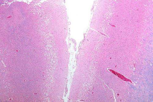
Images of brain after mild stroke predict future risk

A CT scan of the brain within 24 hours of a mild, non-disabling stroke can predict when patients will be at the highest risk of another stroke or when symptoms may worsen, according to new research published in the American Heart Association journal Stroke.
Like stroke, a transient ischemic attack (TIA) is caused by restricted blood supply to the brain. Symptoms may last only a few minutes.
"All patients should get a CT scan of their brain after a TIA or non-disabling stroke," said Jeffrey J. Perry, M.D., M.Sc., co-senior author of the study and associate professor of emergency medicine at the University of Ottawa in Canada. "Images can help healthcare professionals identify patterns of damage associated with different levels of risk for a subsequent stroke or help predict when symptoms may get worse.
Most, but not all, Canadian and U.S. patients with these symptoms undergo CT scanning - an imaging that combines a series of X-ray views to generate cross-sectional images of the brain, he said.
Of 2,028 patients who received CT scans within 24 hours of a TIA or non-disabling stroke, 814 (40.1 percent) had brain damage due to impaired circulation (ischemia).
Compared to patients without ischemia, the probability of another stroke occurring within 90 days of the initial episode was:
- 2.6 times greater if the CT image revealed newly damaged tissue due to poor circulation (acute ischemia);
- 5.35 times greater if tissue was previously damaged (chronic ischemia) in addition to acute ischemia;
- 4.9 times greater if any type of small vessel damage occurred in the brain, such as narrowing of the small vessels (microangiopathy), in addition to acute ischemia;
- 8.04 times greater if acute and chronic ischemia occurred in addition to microangiopathy.
While 3.4 percent of the people in the study group had a subsequent stroke within 90 days, 25 percent of patients with CT scans showing all three types of damage to their brain had strokes.
"During the 90-day period, and also within the first two days after the initial attack, patients did much worse in terms of experiencing a subsequent stroke if they had additional areas of damage along with acute ischemia," said Perry, who is also a senior scientist at the Ottawa Hospital Research Institute.
"These findings should prompt physicians to be more aggressive in managing patients with TIA or non-disabling stroke who are diagnosed with acute ischemia, especially if there is additional chronic ischemia and/or microangiopathy."
Measures to avert a new stroke might include cardiac monitoring or medications to lower blood pressure, treat high cholesterol or prevent blood clots.
The researchers are assessing how to incorporate the study's findings into stroke risk scores that rely on symptoms along with patient factors such as age and the presence of high blood pressure or diabetes.


