A fluorescent molecule selectively labels live neurons in the brain for the first time

Information processing and transmission in the brain hinge on electrical and chemical signals emitted and received by neurons. Now, A*STAR researchers have developed the first small molecule capable of selectively labeling these cells, which is expected to shed light into these complex mechanisms.
Existing neuron labels, such as fluorescent dyes and proteins, exhibit low selectivity or require complicated genetic manipulations that may reduce cell viability. In addition, these systems only work for frozen or fixed brain tissue. In contrast, the new fluorescent probe, known as Neuron Orange (NeuO), exclusively stains live neurons in the presence of other brain cells.
According to team leader Young-Tae Chang from the A*STAR Singapore Bioimaging Consortium, this ability facilitates the real time monitoring of neuron growth and interaction with other cells over extended periods. This provides essential insight on neural development, networking and degeneration.
In a continuous effort to simultaneously visualize the four types of cells coexisting in the brain—neurons, microglia, astrocytes and oligodendrites—Chang's team has searched for probes that can differentiate these cells from each other.
The researchers were able to identify NeuO using high-throughput screening. In this approach, they exposed a library of fluorescent molecules to brain cells cultured in vitro to determine a potential neuron-selective candidate. Next, they systematically modified the functional groups of this candidate and evaluated the impact of these structural changes on its response to enhance its selective staining. This study showed NeuO to be the most neuron-selective stain.
In vitro fluorescence imaging showed that NeuO stained the area surrounding the nuclei and extended to the branched portions of the neurons (see video). Also, the probe did not affect cell survival rate, morphology or function. When combined with a microglia-selective probe for dual imaging, it clearly discriminated a large neuron from other cells.
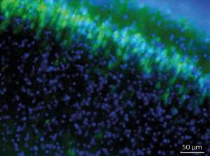
In addition to effectively dying brain slices (see image), NeuO labeled neurons of the hippocampus, cerebellum and spinal cord when administered intravenously into the tail vein of mice. "This means that the dye penetrates the blood–brain barrier and selectively stains neurons in the live brain," explains Chang. "This is quite striking," he adds, noting the potential for use of NeuO in a clinical setting.
The researchers are currently exploring ways to incorporate NeuO into multifunctional probes emitting at different colors to achieve several simultaneous detections. Also, they are improving the properties of this new molecule to expand its potential application to in vivo imaging and drug delivery.
More information: NeuO: a fluorescent chemical probe for live neuron labeling. Angewandte Chemie International Edition 54, 2442–2446 (2015). dx.doi.org/10.1002/anie.201408614


