Clear imaging of pancreatic cells through the development of a novel fluorescent probe

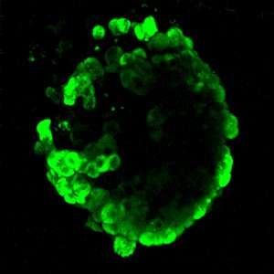
A fluorescent imaging probe developed by A*STAR researchers has allowed scientists for the first time to visualize the detailed three-dimensional distribution of alpha cells in the pancreas of mice1. The probe could enhance diabetes research and shine a light on more effective treatment of the disease.
A key function of the pancreas is to release hormones—namely glucagon and insulin—into the bloodstream to control sugar levels. Glucagon and insulin are stored and released by alpha and beta cells respectively. The alpha and beta cells work so closely together to regulate blood sugar that they form cell clumps known as islets inside the pancreas. Within the islets, glucagon raises the amount of glucose in the bloodstream, and insulin is released when blood glucose levels become too high. Disruption of this regulation is largely responsible for diabetes.
Until now, a suitable fluorescent imaging probe to produce clear pictures of the individual components in live pancreatic islets has eluded scientists.
"It was this gap in bioimaging that we wanted to fill," explains Young-Tae Chang of the A*STAR Singapore Bioimaging Consortium, who led the project. "We wanted to develop a fluorescent probe that could selectively stain glucagon-producing alpha cells, and that was suitable for two-photon microscopy."
This relatively new form of microscopy uses near-infrared light to excite fluorescent dyes. Two photons are absorbed in each excitation, minimizing light-scattering within the tissue. This allows for deeper tissue penetration, more localized and coherent imaging, and quicker observation of cells than is possible via conventional microscopy techniques.
Chang and his team, with collaborators from the National University of Singapore, generated a two-photon fluorescent dye library comprising 80 different compounds. They then used pancreatic cell lines to screen the compounds and identify one that selectively binds to alpha cells and brightly fluoresces. The dye, named Two Photon-alpha (TP-α), performed particularly well when tested on islets isolated from mice, specifically showing alpha cells and their distribution in 3D (see image).
"We have not tested the TP-α probe on human islets yet," states Chang. "If it works, potential applications include diabetes research, as well as possibly islet transplant procedures for diabetes patients. TP-α could be used to monitor the survival and condition of alpha cells, for example."
The team will now focus on testing TP-α in human islets in the laboratory and inside the body. They also hope to extend their two-photon-probe hunt to find one suitable for imaging insulin-producing beta cells.
More information: "Glucagon-secreting alpha cell selective two-photon fluorescent probe TP-α: For live pancreatic islet imaging." Journal of the American Chemical Society 137, 5355–5362 (2015). dx.doi.org/10.1021/ja5115776
Journal information: Journal of the American Chemical Society


