Differences in fat tissues' light reflecting properties make for easy detection

A technique that uses light imaging to monitor how one type of fat tissue is converted to another has been employed by A*STAR researchers to better understand conditions such as diabetes and obesity. The technique could underpin a fast and cost-effective approach to monitoring this conversion.
The two main types of adipose tissue have very different properties: white (WAT) stores excess energy and its accumulation is linked to obesity, while brown (BAT) has immense energy burning capabilities. Some WAT can be converted to BAT-like tissue by a process known as browning which, in rodents at least, has anti-obesity and anti-diabetic functions.
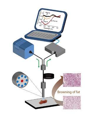
Current methods for monitoring browning are time-consuming and crude. Two teams at the A*STAR Singapore Bioimaging Consortium led by Shigeki Sugii and Malini Olivo collaborated to improve the process. They successfully monitored the browning process using optical spectroscopy techniques—diffuse reflectance spectroscopy (DRS) and multispectral imaging (MSI).
"DRS and MSI made it possible to detect and measure the level of fat browning within minutes," says Sugii. "This is in sharp contrast to traditional analytical methods such as gene expression, protein analysis or histology, which typically take days."
The researchers stimulated browning in WAT from mice and quantified the process using DRS, which detects the pattern of scattered light from a sample that is illuminated with narrow wavelength light. DRS produced distinct spectral "fingerprints" for WAT, browning WAT, and BAT. MSI, which measures reflected light at specific wavelengths, complemented and validated the DRS findings. The researchers further validated these results by comparing them with those from gene and protein analysis methods.
The value in using DRS and MSI for detecting browning comes from the differences in WAT and BAT composition. WAT consists of a single large fat droplet, while BAT comprises many small fat droplets and energy burning machinery (mitochondria). This difference, particularly in composition, means that light imaging techniques can be used to tell them apart. "Optical spectroscopy can fingerprint the intrinsic differences in tissue optical characteristics, thereby differentiating between classical brown and white fat tissue, and white fat during the browning process," explains Olivo.
The researchers anticipate that these techniques can be developed for studying browning in live animals and humans. "These techniques will facilitate the study of associations between obesity and fat browning capacities, and the potential development of therapeutic approaches to enhance browning for tackling obesity," says Sugii.
More information: U. S Dinish et al. Diffuse Optical Spectroscopy and Imaging to Detect and Quantify Adipose Tissue Browning, Scientific Reports (2017). DOI: 10.1038/srep41357
Journal information: Scientific Reports


