Faster image processing for neuroscience

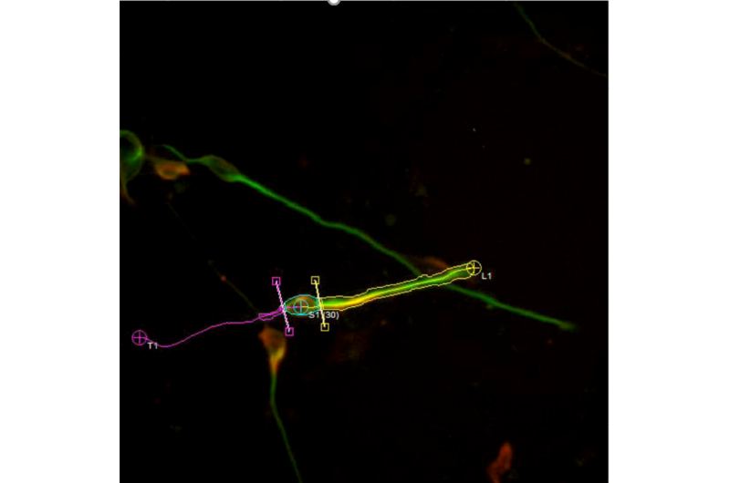
Oak Ridge National Laboratory scientists designed software for St. Jude Children's Research Hospital that significantly sped processing of microscopy images used in brain development research.
The software provided frame-by-frame analysis of video taken of a mouse brain cell in a matter of hours compared with traditional manual techniques that can take weeks. St. Jude's researchers are exploring how neurons migrate in the developing brain to better understand how defects in the process may result in certain disorders such as epilepsy or intellectual disability.
"Automatic processing of the images saves time and provides more data for better statistics in these studies," said ORNL's Ryan Kerekes. Dr. David Solecki of St. Jude's Developmental Neurobiology Department said his collaboration with ORNL has been "indispensable."
The ORNL tools "free up biologists to do what they do best: apply their expert knowledge to analysis tasks," he said. Results of the research were published in Nature Communications.
More information: Niraj Trivedi et al. Drebrin-mediated microtubule–actomyosin coupling steers cerebellar granule neuron nucleokinesis and migration pathway selection, Nature Communications (2017). DOI: 10.1038/ncomms14484


