Identification of the neuronal suppressor of cataplexy, sudden weakening of muscle control in narcolepsy

Narcolepsy is a disorder caused by losing orexin neurons, marked by excessive daytime sleepiness and cataplexy, and sudden weakening of muscle control. Previously, we found two kinds of neurons preventing narcolepsy by receiving information from orexin neurons. Here, researchers have discovered that serotonin neurons inhibit cataplexy by reducing amygdala activity, which controls emotion. The current discovery should lead to understanding of the mechanisms of narcolepsy and possible therapeutic approaches for cataplexy.
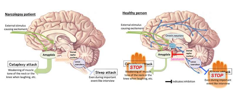
The brain is equipped with a sleep mechanism and a wakefulness mechanism, which are regulated to be on or off in a respective manner. Orexin is important in regulating this switch. If orexin neurons are lost, narcolepsy results.In this sleep disorder, sleep and wakefulness are inadequately switched on and off. The typical symptoms are excessive daytime sleepiness and cataplexy. Cataplexy occurs when the patient is emotionally excited, and in severe cases, they may lose muscle control and fall down. Sleep has two stages, REM and non-REM sleep. Dreams usually occur during REM sleep, when most of the muscles are relaxed (called atonia) in order to prevent the dreamer from unconscious activity. During cataplexy attacks atonia occurs when the patient is awake. The research team previously found two types of neurons preventing narcolepsy by receiving orexin from orexin neurons—the first are noradrenaline neurons in the locus coeruleus of the brain suppressing sleepiness .The other type is serotonin neurons in the dorsal raphe nucleus of the brain, which inhibit cataplexy.
In this study, the international research team led by the researchers of Kanazawa University has discovered that serotonin neurons in the dorsal raphe nucleus inhibit catalepsy by reducing activities of the amygdala that controls emotion.
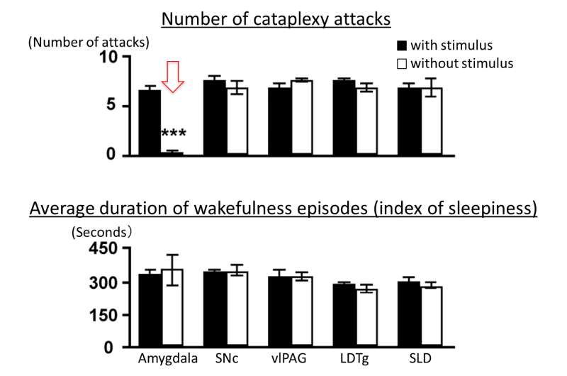
Serotonin neurons in the dorsal raphe nucleus extend projections throughout the brain and send information. In this study, using an optogenetic tool, the team discovered that catalepsy was almost completely inhibited by artificial augmentation of serotonin release induced by selectively stimulating serotonin nerve terminals in the amygdalas of narcolepsy model mice. The same experimental operation in the other brain region that controls REM sleep did not inhibit cataplexy. In addition, the team found that serotonin release reduced amygdala activity. When amygdala activity was artificially reduced in a direct manner, cataplexy was inhibited; when activity in the amygdala was artificially augmented, the frequency of cataplexy attacks increased. Furthermore, the effect of orexin neurons inhibiting cataplexy was found to be abolished when serotonin release was inhibited selectively in the amygdala.
Cataplexy is triggered by sudden emotional excitement of positive valence, such as hearty laughter. This study has revealed that serotonin neurons do not directly suppress muscle control, but inhibit cataplexy by reducing and controlling activities of the amygdala, which is involved in communicating emotional excitement. In fact, it is known that the amygdalas of narcolepsy patients without orexin neurons respond excessively when the patients see, for example, interesting photos. The team believes that the current study has made a big step forward to understanding the whole picture of the narcolepsy mechanism. It is also expected that new therapies will be developed for cataplexy.

More information: Emi Hasegawa et al, Serotonin neurons in the dorsal raphe mediate the anticataplectic action of orexin neurons by reducing amygdala activity, Proceedings of the National Academy of Sciences (2017). DOI: 10.1073/pnas.1614552114


