How Zika virus induces congenital microcephaly

Epidemiological studies show that in utero fetal infection with the Zika virus (ZIKV) may lead to microcephaly, an irreversible congenital malformation of the brain characterized by an incomplete development of the cerebral cortex. However, the mechanism of Zika virus-associated microcephaly remains unclear. An international team of researchers within the European consortium ZIKAlliance (coordinated by Inserm in France) has identified a specific mechanism leading to this microcephaly. Their findings are published this week in Nature Neuroscience.
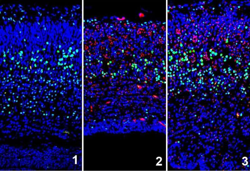
To understand this mechanism, the scientific team led by Dr. Laurent Nguyen of the University of Liège and Prof. Marc Lecuit of the University Paris Descartes combined analysis of human fetuses infected with Zika virus, cultures of human neuronal stem cells and mice embryos. They showed that ZIKV infection of cortical progenitors (stem cells for cortical neurons) controlling neurogenesis triggers stress in the endoplasmic reticulum (where some of the cellular proteins and lipids are synthesized) in the embryonic brain, inducing signals in response to incorrect protein conformation (referred to as "unfolded protein response").
When it reaches the brain, Zika virus infects neuronal stem cells, which will generate fewer neurons, and by inducing chronic stress in the endoplasmic reticulum, it promotes apoptosis, i.e. the early death of these neuronal cells. These two combined mechanisms explain why the cerebral cortex of infected fetuses becomes deficient in neurons and is therefore smaller in size.
"These discoveries demonstrate a hypothesis that we had made following a basic research study we had just carried out in our laboratory, and thus confirm the physiological importance of the unfolded protein response in the control of neurogenesis," says Laurent Nguyen.
Researchers continued their studies on mice by administering inhibitors of protein-folding re-sponse in cortical progenitors and found that this inhibited the development of microcephaly in mice embryos infected with Zika virus. Furthermore, the defects observed are specific to an infection by ZIKV, as other neurotropical viruses of the flavivirus family (West Nile virus, yellow fever,...) did not cause microcephaly, in contrast to Zika virus.
According to Prof. Marc Lecuit, "These results illustrate how studying fundamental biological processes is an essential step in understanding the mechanisms of infections, and lead to novel therapeutic strategies."
More information: Ivan Gladwyn-Ng et al, Stress-induced unfolded protein response contributes to Zika virus–associated microcephaly, Nature Neuroscience (2017). DOI: 10.1038/s41593-017-0038-4


